
Procedure map / Spine
Balloon kyphoplasty
A balloon is inflated inside the vertebral body to create a controlled cavity, which is then filled with bone cement.
Step by step
- 01
Guide wire and cannula
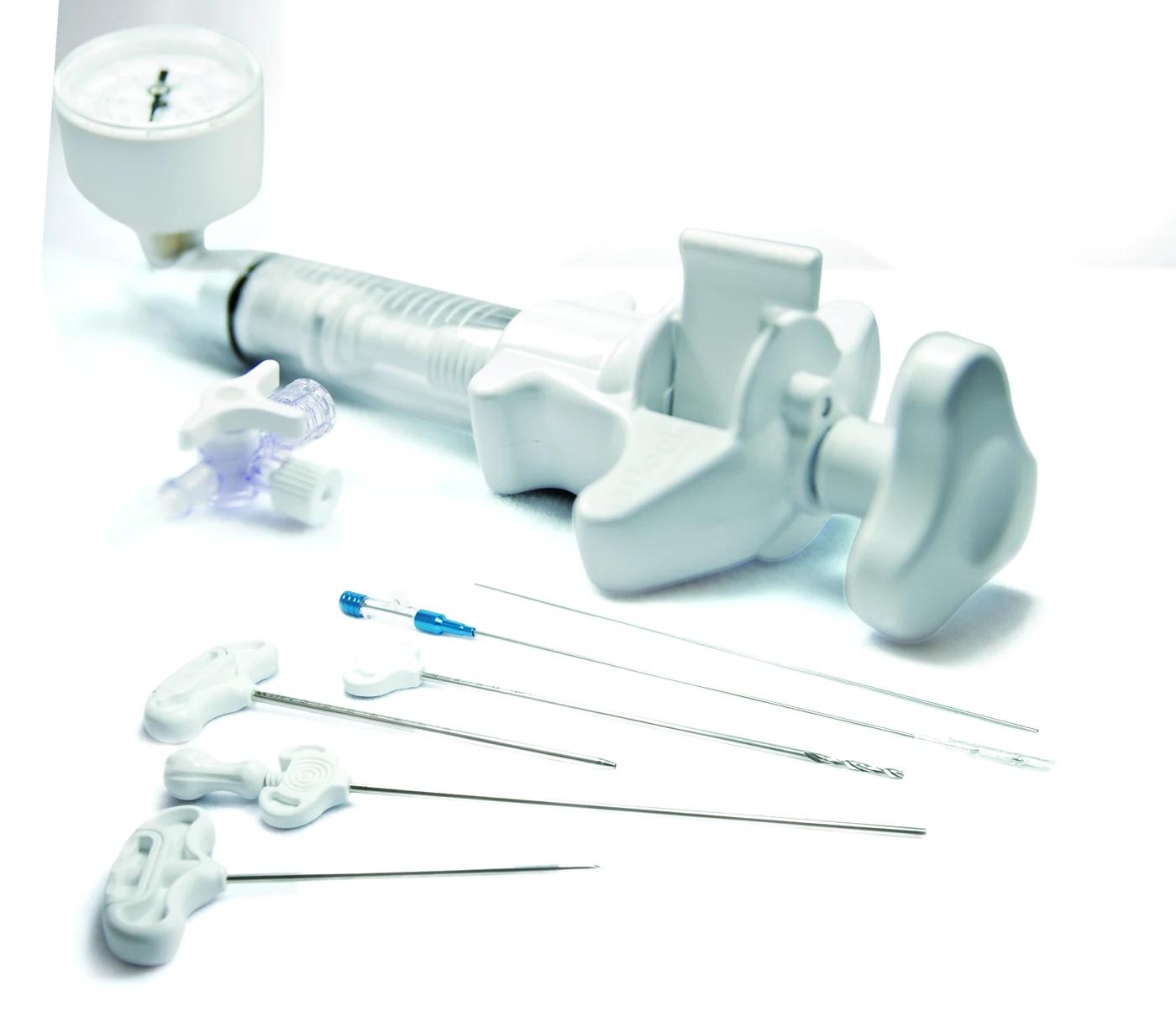
A guide wire is placed through the pedicle; the trocar and cannula are advanced over it into the vertebral body. Usually done bilaterally.
Cannula 178 mm, Ø 4.3 mm
- 02
Opening the balloon path
The bone drill opens a channel in the cancellous bone where the balloon will sit.
Bone drill 220 mm, Ø 3.5 mm
- 03
Balloon inflation
The balloon, filled with contrast, is inflated gradually while pressure and fluoroscopy are watched. Inflation stops once a cavity forms or it reaches the endplates.
Gauge-monitored inflation
- 04
Cement preparation
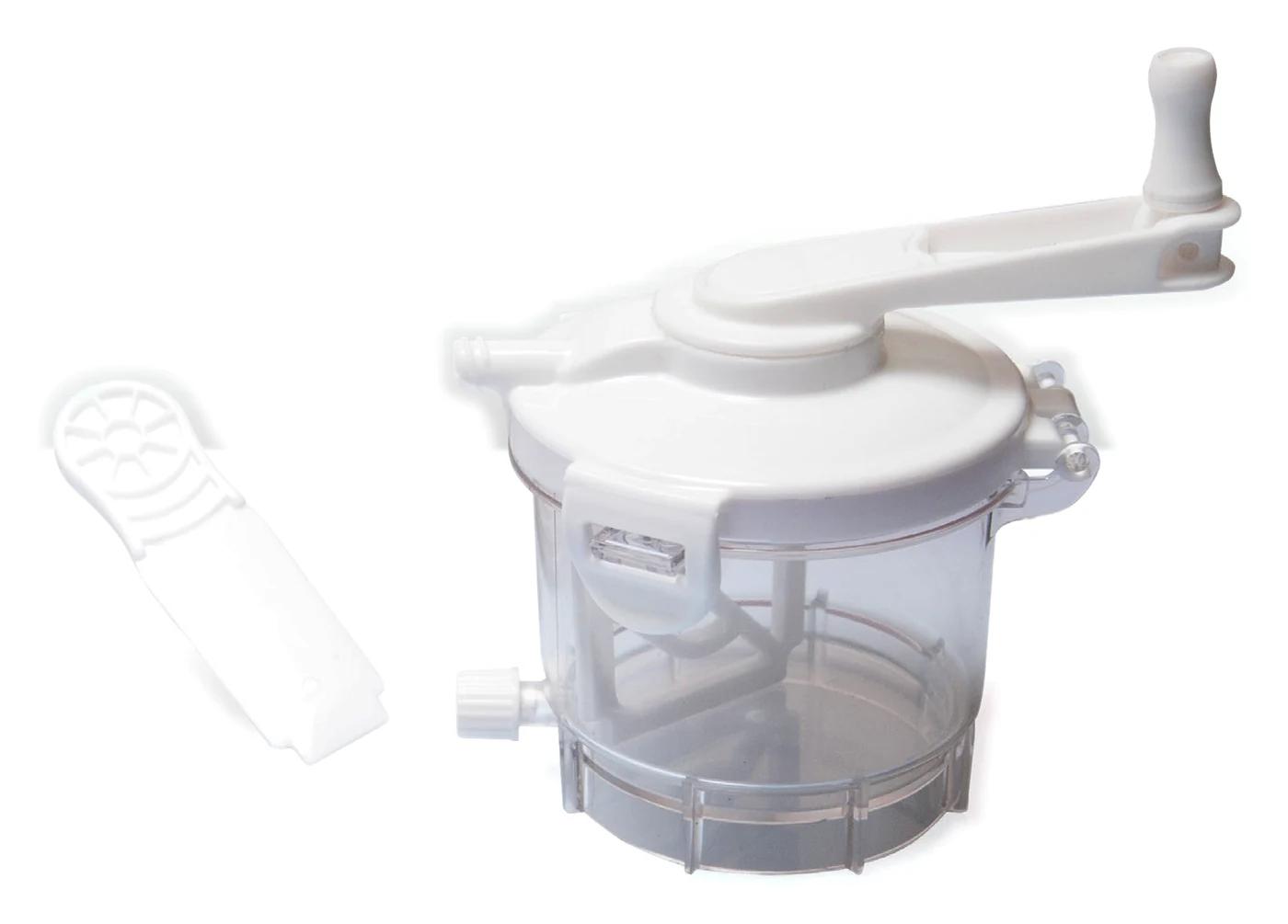
While the balloon is deflated and withdrawn, the cement is prepared under vacuum. High radiopacity keeps the fill clearly visible on screen.
60% ZrO₂
- 05
Filling the cavity
The cavity is filled with the cement fillers and pusher under fluoroscopy, then the cannulas are withdrawn.
Cement filler Ø 3-3.5 mm
A general workflow is shown. Practice varies by physician and patient.