
Procedure map / Pain
Epidural neuroplasty
Epidural adhesions are released with a catheter and medication is delivered directly to the affected nerve root.
Step by step
- 01
Entry
The patient lies prone; under local anaesthesia the epidural space is entered with a needle, usually through the sacral hiatus. The patient stays awake and responsive.
Local anaesthesia, light sedation
- 02
Epidurogram
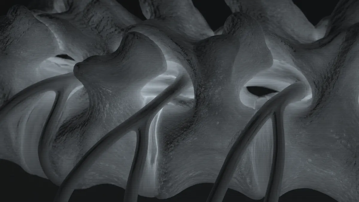
Contrast is injected. Areas where it does not spread show where adhesions (fibrosis) are and which nerve root is affected.
Contrast 5-10 ml
- 03
Steering the catheter
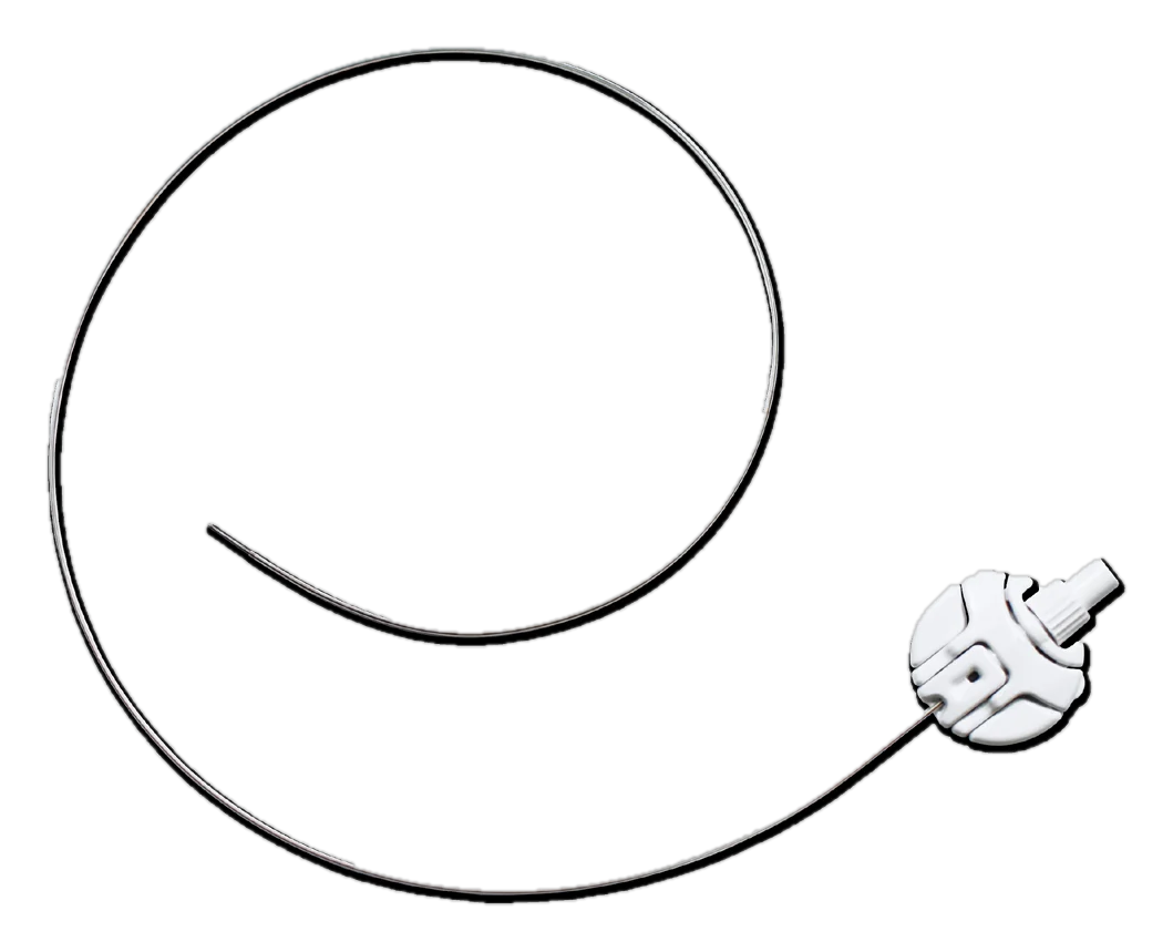
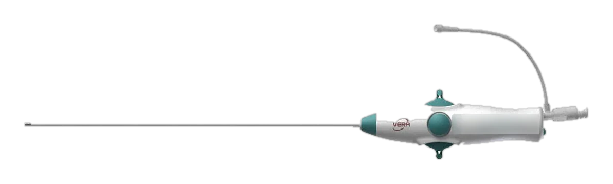
The catheter is passed through the needle and, with its tip bent, advanced under fluoroscopy to the adhesion and the nerve root.
VERA bidirectional tip, PEN spring-guided catheter
- 04
Release and check
The adhesion is released mechanically with the catheter tip. Repeat contrast spreading along the nerve root shows the release is adequate.
Radiopaque, atraumatic tip
- 05
Medication delivery
Following the physician's protocol, agents such as hyaluronidase, local anaesthetic and corticosteroid are delivered to the target in sequence. The catheter is withdrawn.
Medication is given through the catheter's injection port
A general workflow is shown. Practice varies by physician and patient.